Electron- and proton transport through multi-redox center proteins incorporated in biomimetic membrane systems is investigated by surface-enhanced IR absorption spectroscopy (SEIRAS), fast-scan voltammetry, fluorescence and UV-Vis spectroscopy.
Active transport is initiated by direct electron transfer and alternatively by photo-excitation. Model calculations are employed for mechanistic studies.
Proteins Incorporated in Biomimetic Membrane Systems
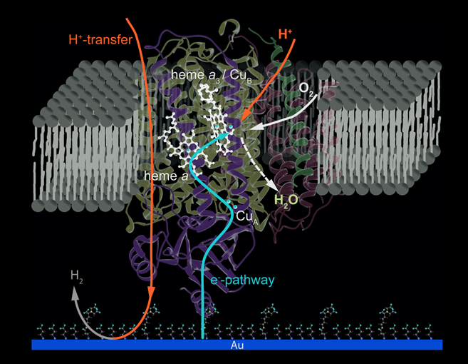
Cytochrome c Oxidase from R. sphaeroides, immobilized via his-tag engineered to SU II reconstituted in a protein-tethered bilayer lipid membrane (ptBLM) Electrochemical excitation by direct electron transfer
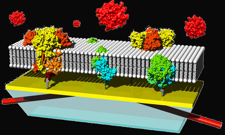
RC and bc1 proteins immobilized on the NTA-modified planar surface = (gold) of an ATR crystal (transparent blue) (B). The cytochrome c binding site of = both proteins is located on the outer side of the lipid layer. Rhodobacter sphaeroides RCs (green) with a genetically engineered 7-his-tag (dark = green) at the C-terminus of the M subunit, and bc1 complexes (orange) poly-his-tagged (dark orange) on the C-terminus of the cyt b subunit. Cytochrome c is depicted in red.
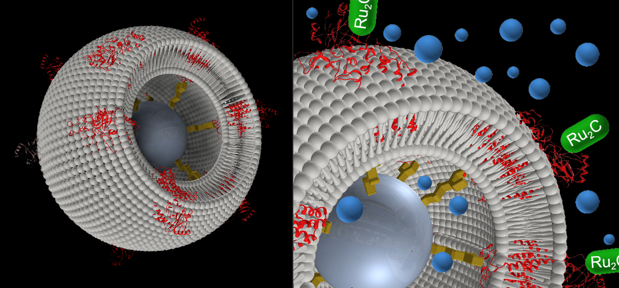
Proteo-Lipobeads encapsulating cytochrome c oxidase ( CcO) from P. denitrificans with a his-tag engineered to SU I. Excitation by flash-induced electron transport via Ru complexes.